08 Sep 2023 The Role of Ultrasound in the Diagnosis and Treatment of Hernias
In the field of medicine, advancements are occurring simultaneously across almost every specialty, with interactions and solutions tailored to specific needs. Technology plays a significant role, particularly in advanced imaging technologies. Among the most well-known are ultrasound, computed tomography (CT), and magnetic resonance imaging (MRI). In this blog post, I will emphasize the importance of ultrasound in hernia surgery.
What is Ultrasound?
Ultrasound involves sending sound waves at a frequency beyond human hearing to a specific area of the body, recording their reflections in real time to create an image. For instance, while the sound waves from an ultrasound device pass effectively through solid organs and soft tissues, they do not penetrate air-filled organs like the digestive system. Additionally, bones do not transmit ultrasound waves, allowing organs surrounding bones to be examined using this technique.
The closer the probe (the part of the ultrasound device that makes contact with the body) is to the organ being imaged, the higher the quality of the image. To overcome the obstacle of air, systems such as endo-ultrasound devices that can pass through the channels of endoscopes have been developed for purposes like performing ultrasounds inside the digestive tract. Similarly, ultrasound probes compatible with both open and laparoscopic systems have been produced to accurately determine the borders of masses in the liver during surgery. The principle remains the same: being as close as possible to the organ to be imaged.
Ultrasound offers various advantages: it is practical, fast, and efficient. It is readily available and easily accessible. Imaging does not take much time and can be conducted rapidly. Ultrasound devices are much cheaper compared to CT and MRI machines. They can be transported anywhere, greatly expanding their range of applications. They can be used in the operating room, patient rooms, ambulances, and even outdoors.
In hernia surgery, ultrasound provides valuable assistance in diagnosis and treatment.
Diagnostic Ultrasound in Hernia Surgery
Let’s start with diagnosis: inguinal and abdominal wall hernias can be easily diagnosed using ultrasound. Additionally, it is highly effective in measuring the dimensions of the hernia, identifying adjacent structures, determining the condition of muscles and soft tissues, measuring diastasis, and revealing hidden hernias. I’d like to provide a few examples and touch on some debated topics regarding its use.
The Contralateral Hernia Conundrum!
Knowing the condition of the contralateral side is crucial in cases of unilateral inguinal hernia. With the increasing use of laparoscopic repair techniques and the adoption of ultrasound, the incidence of bilateral inguinal hernias has dramatically risen, with reports ranging from 5-60%. It has been observed that up to 30% of patients with a history of inguinal hernia surgery can develop a hernia on the contralateral side over time. This underscores the necessity for a comprehensive evaluation of both sides of the inguinal region. Although physically examining both sides seems to be the most suitable approach for hernia diagnosis, the use of ultrasound for examining the inguinal region nowadays reveals an excess diagnosis of inguinal hernias. Consequently, patients diagnosed with bilateral inguinal hernias are more frequently encountered, even if the initial symptoms are unilateral.
Most patients diagnosed with bilateral inguinal hernias only experience symptoms on one side. Performing a simultaneous repair of both hernias in the same operation, under a single anesthesia, through the same incisions, with a short recovery period and low complication rates, are the fundamental advantages of the laparo-endoscopic approach. On the other hand, bilateral repairs are associated with higher costs due to longer operating times and the use of two meshes compared to unilateral repairs. There is potentially preventable morbidity on the asymptomatic side, with reported chronic pain rates of up to 10-15% in scientific publications. Therefore, making a decision in cases of bilateral inguinal hernias, where one side is asymptomatic, is truly challenging.
A comprehensive preoperative assessment of the contralateral side, primarily through physical examination and dynamic ultrasound, should be routinely performed, and if an asymptomatic hernia is detected on the contralateral side, bilateral repair should be considered. But this decision should be discussed with the patient and he/she should be included in the decision making process.
If a patient presents with groin pain and no distinct bulging is observed, and a hernia cannot be diagnosed through physical examination, ultrasound becomes an essential tool for diagnosis. In this case, a dynamic ultrasound should be conducted. The patient should be induced to strain or cough, a maneuver known as the Valsalva maneuver, to attempt to reveal the hernia. If a hernia cannot be detected through ultrasound and it is necessary to identify the cause of the pain, MRI imaging must be conducted. Blindly performing surgery claiming that the pain is due to a hernia should never be done. It is highly likely that the patient’s pain will persist after the operation. There can be many reasons for the pain besides a hernia!
What we want to see in a groin hernia ultrasound
Ultrasound helps us in preoperative planning. We want to know if the hernia is directly from the posterior wall of the groin, or if it is an indirect type coming through the inguinal ring. We want to know if there is an accompanying lipoma in the canal. We want to know if there is a hernia in the femoral canal. These are what we as hernia surgeons want to see in an ultrasound report. Otherwise – as we often see in reports – the size of the hernia is not very important. What matters is that there is a hernia; the size of the hernia hole in the groin doesn’t matter. Whether it is 5 mm or 5 cm, the repair will be done with the same standard approach using a mesh determined in the guidelines.
Femoral hernia on ultrasound
Missed femoral hernias are the cause of about 10% of recurrent groin hernia repairs done with an open technique. We see this most commonly in women. Therefore, groin pain, groin hernia, and femoral hernia in women show differences from men and should be carefully examined. This is emphasized in the Lichtenstein technique, which is the most popular technique for open hernia repair. During surgery, the groin floor should be opened to check the femoral canal. If there is a hernia, the mesh should be appropriately cut and sewn to Cooper’s ligament. However, it is useful to know before the repair. If a femoral hernia is known to be present, it is better to repair it laparoscopically. We especially recommend laparoscopic repair of groin hernias in women.
Ultrasound in recurrent groin hernia
Recurrent groin hernia is a common occurrence. While the use of meshes significantly reduces recurrences, worldwide averages remain at unacceptable rates like 7-14%. The goal is 0.5%! The diagnosis is of course made through physical examination, but it may not always manifest as a clearly visible bulging. Sometimes burning or pain may be signs of a recurrence. Whether there is bulging or not, ultrasound is the first imaging method to be used. In addition to making the diagnosis, it can reveal the condition of the tissues in the groin and the status of the previously placed mesh. However, if this is not sufficient, CT scan is the next option.
Ultrasound in the diagnosis of abdominal wall hernias
Ultrasound is sufficient for primary hernias of the anterior abdominal wall, such as umbilical hernias, epigastric hernias, and Spigelian hernias, and provides valuable information. Surgical hernias (incisional hernias) have distinctive features and for diagnosis and surgical planning, CT is the preferred imaging method. Again, we prefer CT in recurrent abdominal wall primary hernias.
Ultrasonography in umbilical hernias
Ultrasound provides valuable information in umbilical hernias: the size of the hernia, the organs entering or trapped within it, the presence of accompanying rectus diastasis (I will discuss this separately), and the presence of accompanying epigastric hernias. Evaluating these together directly affects the success of umbilical hernia repair.
Sometimes epigastric hernias accompany umbilical hernias. Epigastric hernias generally do not cause symptoms but can grow over time and cause pain. If we detect this with ultrasound, we include it in the repair.
The size of the hole in the umbilical hernias (usually circular in the navel) is important. For hernias larger than 2 cm, we prefer laparoscopic techniques. In smaller hernias and non-obese patients, repair through a cut in the navel hole is possible. Ultrasound allows us to measure this without any problems.
Ultrasound in rectus diastasis
The importance of rectus diastasis has been understood in recent years. When repairing midline abdominal hernias, rectus diastasis is now also included in the repair. It is said to contribute to preventing recurrences. We still don’t have enough data on this! The diameter of the diastasis is important. We include it in the repair for diastasis exceeding 3 cm. Ultrasound provides us with information about the diameter, length of the diastasis, and the condition of the rectus muscles. These help us determine the repair technique. For example, in diastasis wider than 5 cm, considering the option of using a mesh is necessary. Otherwise, recurrences may increase. Sometimes there are very large diastases that can negatively affect the patient’s abdominal wall functions, respiration, and cause back pain. In this group of patients, techniques involving component separation may be needed.
The therapeutic application of ultrasound in hernia surgery
Ultrasonography plays two crucial roles in hernia surgery: providing minimally invasive interventions for post-operative complications and performing nerve blocks. Hernia surgery is typically performed in soft tissues, involving the creation of layers and spaces to repair and place a mesh. In these areas, postoperative complications such as hematoma due to bleeding, seroma caused by serous fluid accumulation, and more severe issues like infection and abscess formation may occur. Effective drainage and placement of drains become necessary for these accumulations. These procedures are best performed under the guidance of ultrasound, ensuring safe and effective outcomes.
Another application is the preoperative identification of anatomically significant landmarks using ultrasound for surgical planning. Especially during eTEP-ventral procedures, determining the location of the linea semilunaris instead of relying on palpation, and delineating the dimensions and boundaries of the hernia on the skin surface, greatly contributes to the surgeon’s spatial orientation.
Chronic pain after inguinal hernia surgery is a significant issue, most commonly attributed to nerve damage and presenting as neuropathic type pain. Ultrasonography is at the intersection of diagnosis and treatment for this condition. The nerves most frequently affected by chronic pain after inguinal hernia surgery are the Ilioinguinal, Iliohypogastric, and Genitofemoral nerves. With ultrasound, we can visualize these nerves with high precision and simultaneously perform temporary or permanent blocks. Temporary blocks serve a diagnostic purpose. If the pain subsides after a temporary block is performed on a nerve suspected to be damaged, it leads to the diagnosis that the pain stems from the injury to that nerve, thus informing the treatment plan.
Another significant application is pain blockage. In cases of large hernia surgeries where abdominal wall reconstruction is performed, a Transversus Abdominis Plane (TAP) block, involving the administration of local anesthetic to the transversus abdominis muscles on both sides, significantly contributes to reducing post-operative pain. The innervation of the abdominal wall muscles comes from the intercostal nerves, which progress between the internal oblique (IO) and transversus abdominis (TA) muscles towards the rectus muscles, along with associated arteries and veins. This structure is referred to as the neurovascular bundle, and at this point, they are called the rectus perforators.
TAP block can be quickly performed under ultrasound guidance after the patient has been sedated before the surgery starts. Since it is performed before the surgery, it is referred to as preemptive anesthesia and is more effective than local anesthesia administered after the surgery. Bupivacaine is used for local blocks, and its effect lasts for 6-8 hours. In the United States, a liposome-encapsulated form of bupivacaine, marketed as Exparel®, is used, and it remains effective for 72 hours. However, this drug, which is quite expensive, has not yet been licensed in our country. Additionally, the rectus sheath block under ultrasound guidance is a highly effective method.
Finally, I will mention a blockage achieved through botox injection, which involves temporary denervation of the lateral muscle group. Temporarily blocking the trio of muscles on the lateral side (EO, IO, and TA) under ultrasound guidance helps increase intra-abdominal volume and aids in the tension-free closure of hernia defects, especially in large hernias. This procedure is a significant step in abdominal wall reconstruction and is becoming increasingly common. We also apply this approach in our clinic.
Looking ahead: ultrasound now fits in your pocket!

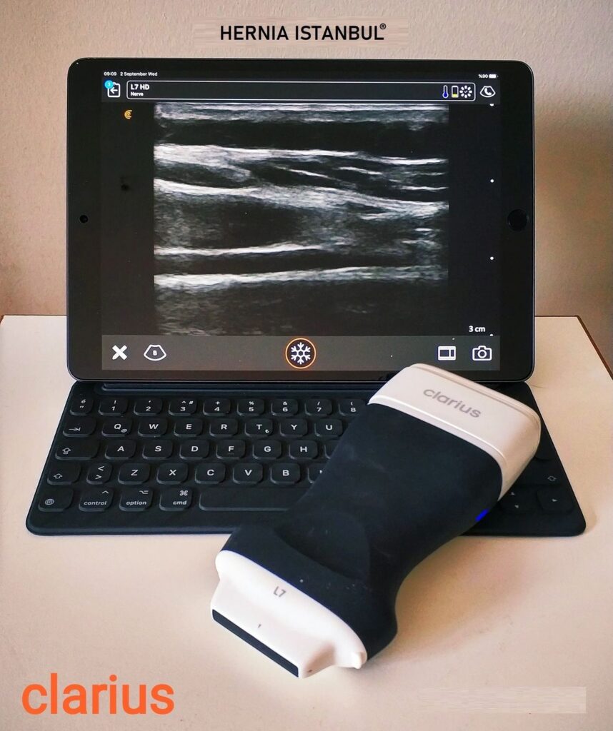
Ultrasound devices, which used to be the size of a refrigerator, have now shrunk to the level of a laptop. There are even probes that work with iPads and smartphones. However, the pinnacle in this regard is ultrasound systems with wireless probes. A probe the size of a cell phone connects wirelessly to the ultrasound software on an iPad using its own Wi-Fi technology, transmitting real-time high-quality images. Ultrasound now fits in your pocket! Thanks to the software, you can transmit the image in real time to anyone anywhere in the world and receive their input and feedback. The scope of application for such technologies is expanding. In line with these developments, we acquired our ultrasound device to address the identified need in hernia surgery. After extensive research and consultations, we purchased the wireless Clarius® ultrasound device, manufactured in Canada. We use it frequently in our daily practice and are very satisfied with its performance.